
The Center for the Advancement of Multicultural Perspectives on Science is pleased to host the CAMPOS Research Colloquia. The Research Colloquia showcase the fantastic research done by CAMPOS scholars and aims to continue building a diverse scientific community in STEM at UC Davis.
There are currently 50 CAMPOS Faculty Scholars at UC Davis, with appointments spanning 34 departments in 8 colleges and schools.
The CAMPOS Research Colloquia are organized by Verónica Martínez Cerdeño.
Previous Colloquiums
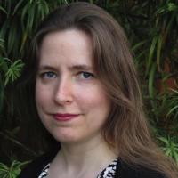
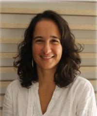
Marie Heffern
Associate Professor, Chemistry
"Probing Metal Dynamics in the Extracellular Environment for Disease Biomarkers"

The context in which a metal resides within a biological environment significantly influences its activity in function. Recent years have seen a rise in tools for monitoring metal ions and have illuminated the diversity in metal speciation in biology, but many of these tools are focused on probing metals within cells and tissues. The state-of-the-art methods for assessing metal status in extracellular fluids such as blood plasma focus either on absolute quantitation or evaluate a limited number of metal-containing species. While these methods have offered important insight into extreme cases of metal deficiency in overload, subtle imbalances are more challenging to diagnose and understand. This talk will describe our efforts to expand and elucidate metal speciation and its dynamics in the extracellular space, specifically in the blood plasma and the interstitial space. We will discuss molecular-level investigations of extracellular biomolecules and their interactions with metal ions, the development of tools to selectively probe extracellular metal biology, and the impacts our research can have in discovering biomarkers that link metal micronutrient homeostasis, diet, and metabolic disorders.
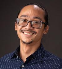
Marco Gonzalez Contreras

Associate Professor, Neurology
"Molecular alterations leading to seizures"
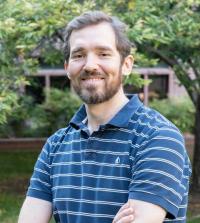
Jesús M. Velázquez
"Advancing a Sustainable Future for All: Materials Design and Energy Conversion Through the Lens of Chemistry"
In this talk, I will share my professional journey, from my undergraduate years in Puerto Rico to my faculty position at UC Davis. I will then discuss my team’s work on developing solid-state synthetic pathways using earth-abundant materials to address energy demands and carbon emissions. We explore structure–function relationships in multinary chalcogenides, with molybdenum sulfides showing promise as CO₂ reduction catalysts. Additionally, I will present findings on electrosynthesizing metal oxalate cements from CO₂. Lastly, I will highlight programs we have designed to support first-generation, low-income students through intervention classes and undergraduate research opportunities.
Martine Therrien
Assistant Professor
Molecular and Cellular Biology
Leveraging Stem Cell Models to Perform Inclusive Functional Genomic Studies
Despite significant advancements in biomedical research, translating these innovations into universal clinical applications remains a complex task. One of the foremost challenges is the biological diversity among patients, including genetic and environmental factors, which can result in varying treatment responses.
Induced pluripotent stem cell models offer unique insights into cellular processes by recreating the genetic landscape of patients. The ability of these models to differentiate into multiple cell types makes them invaluable for understanding disease mechanisms. My talk will discuss how we leverage functional genomic tools to study Alzheimer’s disease across dozens of patient cell lines simultaneously and how it could be applied to other diseases.
Felicity Muth
Assistant Professor
Neurobiology, Physiology and Behavior
How do animals think? Some insights from bumblebees.
February 20, 2025: Animals use their cognition (perception, learning and decision-making) to navigate their worlds, making complex decisions. Many of these processes are shared amongst humans and non-human animals. Here I draw on a few topics from our research to highlight ways that bees (and perhaps other animals too) think about things. In addition, I discuss some recent outreach events from our lab and efforts to spread knowledge about native bees.
Read an article about Muth's funding from The National Geographic Society for her project, "Ecology Shaping Cognition: An Exploration with Wild Bees."
Muth is also a children's book author! Learn more about: "Am I even a bee?"
Natalia Caporale
Associate Professor of Teaching
Neurobiology, Physiology and Behavior
Asset-based Frameworks to Promote Student Success in STEM
February 5, 2025: Efforts aimed at promoting the retention and success of students from minoritized populations in STEM have traditionally followed a student-deficit approach, where equity disparities are seen as a representation of students' deficits such as lack of motivation, lack of preparation, lack of career readiness, etc. This perspective ignores the role of the systemic structural barriers and issues present in Institutions of Higher Education in creating and perpetuating the observed disparities in student outcomes and experiences. In addition to the damage that the deficit perspective does to students, ignoring the role of these structures prevents engagement in discussions and institutional transformation needed in order to address the existing inequities. In this talk, I will share perspectives and findings from my research group in the context of three mixed methods studies that use student asset-based frameworks to examine student experiences and outcomes: (1) Experiences and statistics of Latinx students on Academic Probation, (2) Presence of systemic gender gaps in upper division biology courses across several R1 institutions, (3) Ethnic Studies courses as support mechanisms that promote the success of all students at UC Davis.
Maike Sonnewald
Assistant Professor, Computer Science
Trustworthy AI for Ocean Dynamics: A Semantics-Driven Approach to Understanding Ocean Circulation
January 22, 2025: Artificial Intelligence offers transformative potential in climate science, particularly in understanding complex and observationally sparse regions like the Southern Ocean encircling Antarctica. However, this potential comes with the dual challenge of making AI algorithms that are fit-for-purpose and managing limitations, such as its ‘black box’ aspect. In this talk, I present a methodology grounded in AI-semantics for new knowledge. First, an unsupervised AI framework that centers validation and uncertainty quantification using manifold mapping and clustering identifies distinct dynamical regimes within the Southern Ocean: areas of sparsity in the equations governing the circulation. These regimes provide new, physically meaningful insights into ocean circulation. Second, emphasizing the need to validate neural network predictions critically, we use a semantics-driven neural network approach that ensures predictions are grounded in geophysical fluid dynamics and subsequently demonstrate how this can be used to gain new knowledge. By using neural networks enhanced with explainable AI, we track these dynamical regimes using sparse available data, and use the neural network’s changes in input relevance to guide hypothesis generation and testing. We reveal how fundamental shifts in the drivers of Southern Ocean circulation are governed by climate change. This work serves both as an exploration of AI's promise and a blueprint for responsibly navigating the challenges it presents in climate science, discussing standards for reliable, transparent, and hypothesis-driven AI applications.
Jairo Fúquene Patiño
Assistant Professor, Statistics
Bayesian Methods to Estimate the Completeness of Death Registration

January 8, 2025–Civil registration and vital statistics (CRVS) systems should be the primary source of mortality data for governments. Accurate and timely measurement of the completeness of death registration helps inform interventions to improve CRVS systems and also generate reliable mortality indicators. In this work, we propose the use of Bayesian models to estimate the completeness of death registration at global, national and subnational levels. We propose suitable Markov chain Monte Carlo algorithms to measure the uncertainty of the predictive completeness at the different levels and study the theoretical properties of the Bayesian models. The use of our approach can allow institutions to improve the model parameter estimates and prediction of completeness of death registration. Our new models are based on a dataset updated based on 120 countries and 2,748 country-years. To illustrate the effectiveness of our proposal at national and subnational levels, we consider the completeness of death registration in a low-income country as our comparator dataset. This talk is based on joint work with Tim Adair, Melbourne School of Population and Global Health, The University of Melbourne.
Jasquelin Peña
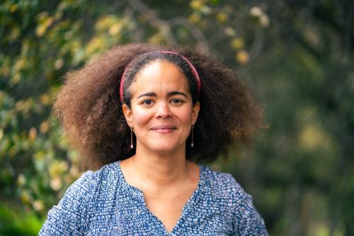
Professor, Civil & Environmental Engineering
Faculty Scientist, Lawrence Berkeley National Laboratory
Building Climate Resilience in Post-fire Landscapes through Geochemical Observations and Community Engagement
December 4, 2024–Wildfires are raising unprecedented challenges in the western United States. Its effects are manifest as both abrupt and long-term changes in ecosystem function. The catastrophic fire seasons across the western U.S. in 2020 and 2021 provided a preview of the magnitude of environmental impact and cost that will be associated with future fire events. One key feature of post-fire landscapes is the transformation of above-ground biomass and litter into a surface layer of ash. This ash layer, which is commonly enriched in metals and pyrogenic carbon, can impact the retention or loss of organic matter, nutrients and trace elements from post-fire landscapes. These impacts will be manifest not only in changes in soil biogeochemistry, but also as changes in surface runoff and infiltration that also impact stream chemistry. In this talk, I will share focus on our analyses of samples from the Glass Fire (2020) and the Caldor File (2021) in order to develop process-level understanding of the response of mountain ecosystems to wildfire and inform resilience building.
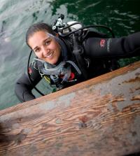
Verónica L. Morales
Associate Professor, Civil & Environmental Engineering
Transport and Reactions in Structurally Dynamic Soils and Rocks: From Dissolution to Bioclogging

October 23, 2024–Reactive transport in porous media is of central importance to many environmental applications spanning groundwater contamination, oil recovery, and geotechnics. Accurate descriptions of water flow, chemical transport, and biochemical reactions in soils and rocks warrant a firm understanding of the pore-scale dynamics at play. The underlying structure of porous media impacts the uniformity of fluid flow, the resulting chemical gradients, and consequently how much reaction will be achieved. The fact that some reactions act to alter the pore structure complicates things further. This talk will investigate two processes that modify the pore structure as a consequence of a reaction: dissolution and bioclogging. Through a deeper understanding of the pore structural changes, evolving flow, and mixing dynamics, it is possible to infer when and how the system can up/down regulate the overall reactivity in time to better target the desired engineering application.
Alan Lombard

Assistant Professor, Biochemistry and Molecular Medicine and Urology
Characterization of PARP Inhibitor Response in Prostate Tumor Cells Reveals Drug Tolerant Persistent Phenotype
October 23, 2024 – PARP inhibition is significantly improving how we manage advanced prostate cancer. Despite this progress, resistance and progression remain inevitable. Our findings reveal that response to PARP inhibition may be characterized as drug tolerant persistence, which could be a path toward development of treatment insensitivity. We seek to understand and target the drug tolerant persistent phenotype to improve treatment efficacy and improve patient outcomes.
Crystal Rogers
Associate Professor, Anatomy, Physiology and Cell Biology
School of Veterinary Medicine
Neural crest cells are the best cells
May 15, 2024–Who am I if I am not a developmental biologist? I am a first-generation college graduate and a scientist who is passionate about my research field. The work in my lab focuses on understanding the molecular and cellular mechanisms that drive some of the earliest processes in vertebrate embryonic development. How do cells choose their fate? How do they migrate? How do tissues form? How are faces made? In this CAMPOS colloquium talk, I will speak about my path in science and how I discovered the field of developmental biology. I will also share some exciting results, gathered by the amazing scientists in my lab, about how neural crest cells form and differentiate during embryogenesis and what happens if those processes are perturbed.
Fernanda Ferreira
Professor, Psychology
Karina Tachihara
Post-doctoral Scholar, Ferreira Lab
“Multi-Utterance Language Production as Foraging”
April 17, 2024–How do speakers plan complex descriptions and then execute those plans? In this work, we attempt to answer this question by asking subjects to describe complex visual scenes while simultaneously monitoring their eye movements. We posit that speakers begin planning by organizing the scene into meaningful clusters or groupings of objects centered around both physical and semantic distance. Speakers then describe the scene cluster by cluster, exploiting the content available at one cluster before moving on to another. To test these ideas, in a preregistered study 30 participants described 30 indoor and outdoor scenes while their eye movements and speech were recorded. Physical distance was calculated by identifying the centroid point of each object and then computing the Euclidean distance between centroid points for every object pair. Semantic distance was calculated using ConceptNet Numberbatch to obtain the semantic similarity between object labels. A clustering algorithm was then applied to establish the appropriate number of clusters per scene and to assign objects to each physical or semantic cluster. We observed that, consistent with our hypothesis, objects separated by shorter physical distances and objects that are semantically more similar were discussed in closer temporal proximity in the verbal descriptions. In addition, word productions that involved jumping from one predefined cluster to another took longer to initiate than those associated with the same cluster. We conclude that speakers solve the linearization problem by establishing physical and semantic clusters and exploiting the information contained at each one. This approach treats multi-utterance language production as a type of foraging behavior, where people balance exploration and exploitation by locating an object cluster, exploiting the contents of that cluster, and exploring the environment for new clusters once the current one is depleted.
Alexander Gamero-Garrido
Assistant Professor, Computer Science
Interactions Between the Internet & Policy: Impacts on Security, Privacy & Resilience
April 3, 2024–The Internet is an essential service. Yet, its connecting infrastructure remains poorly understood, particularly at the country level. This obscurity hampers efforts to evaluate both the resilience of the Internet as well as the security and privacy of digital communications. In this talk, I will present research approaches that I have developed at the intersection of Internet technologies and public policy. My methods empirically and quantitatively assess the resilience, privacy and security of the Internet in many jurisdictions worldwide. The focus of this presentation is on three key research questions: (1) Which countries have the most centralized networks? Centralized infrastructure is problematic as it is less resilient to observation and disruption. (2) Which countries’ networks are dominated by state-owned Internet providers? These countries are exposed to government surveillance and the infringement of civil liberties, particularly in autocracies. (3) Do privacy laws impact the location of web servers? If servers are located in jurisdictions with weaker privacy protections than those of the users’ home nations, users may thus be exposed to unlawful surveillance by advertisers and foreign governments. My studies answer these questions by collecting empirical data on the Internet at global scale and developing novel data-analysis frameworks, along with linking Internet features to policies and individual harms. A key result of this research is the identification of countries that are most exposed to Internet surveillance, especially by governments and advertisers. My presentation concludes with future directions on the impacts of Internet technologies and related policy on historically marginalized communities worldwide.
Marco Gonzalez
Associate Professor, Neurology
Proteolysis and epileptogenesis
March 20, 2024–Epilepsy is a brain disorder characterized by the occurrence of seizures as a reflection of abnormal electrical activity in the brain. The excessive electrical activity in neurons of the brain may be the result of a brain injury or a genetic mutation. The process of brain transformation due to a brain injury or genetic mutation leading to the occurrence of spontaneous recurrent seizures is known as epileptogenesis. We will explore some of the mechanisms promoting the transformation of a normal brain into an epileptic one. The focus will be on the use of rodent models of epilepsy to study the role of proteolysis on the regulation of neuronal proteins during epileptogenesis.
Luis Diaz-Garcia
Assistant Professor, Viticulture and Enology
“Grapevine Rootstock Breeding at UC Davis”
March 6, 2024–Most of the worldwide grape production relies on rootstocks tolerant to biotic and abiotic stresses in the soil. In addition, rootstocks enable the cultivation of high-quality grapes across diverse soil conditions and allow growers to manage productivity. Many Vitis species have potential as rootstocks; however, about 90% of the grapevines worldwide are grafted onto fewer than 10 commercial rootstocks, most of which originate from only three species. During this talk, I will discuss our efforts in grape breeding at UC Davis aimed at integrating greater genetic diversity into the development of new, more resilient rootstocks. I will also cover the various approaches, including phenomics and genomics, that are instrumental in accelerating our breeding pipelines.
Maciel Hernández
Assistant Professor, Human Ecology
"A person-centered examination of culturally-relevant school practices and academic functioning in adolescence"
Schools are sites where youth might have a range of opportunities to be holistically supported through culturally-relevant practices. Using a person-centered approach, we examined the presence of profiles of culturally-relevant practices in schools among adolescents (N = 717; 50% girls; Mage = 13.73 years) from the U.S. Southwest. Four culturally-relevant support profiles were identified. A high culturally-relevant support profile, with relatively high culturally-relevant practice experiences, had consistently higher emotional and behavioral engagement scores than culturally-devoid support, low culturally-relevant support, and moderate culturally-relevant support profiles, which had low to average culturally-relevant practice experiences. Emotional and behavioral engagement were also higher in the moderate and low culturally-relevant support profiles compared to the culturally-devoid support profile, which had relatively moderate levels of teacher support but very low promotion of cultural competence and critical consciousness socialization. Fewer but some similar patterns emerged for school attendance and GPA. The results emphasize the associations between profiles of culturally-relevant support in school and various indicators of academic functioning.
Juliana Leite Nobrega de Moura Bell
Associate Professor, Food Science and Technology
“Boosting almond milk: sustainable strategies for better nutrition and taste”
February 7, 2024–Despite being dominant in the US plant-based milk market, almond milk faces challenges like low protein content, gritty texture, and particle separation due to the current solid dispersion manufacturing process. This challenge has prompted our laboratory to explore the use of a sustainable water and enzyme-based co-extraction approach that allows for replacing the dispersion approach by the simultaneous extraction of proteins and lipids. By judiciously optimizing important extraction parameters such as particle size, roasting level, extraction pH, and enzyme concentration, with respect to composition, yields, physicochemical and sensory properties, almond milk formulations with enhanced nutritional composition (i.e., higher protein content) and high consumer acceptance were developed.
James Letts
Associate Professor, Molecular and Cellular Biology
“Emerging understanding of eukaryotic respiratory diversity”
January 24, 2024–Respiration is a core biological energy-converting process whose last steps are carried out by a chain of multi-subunit complexes in the inner mitochondrial membrane. Complex I is the main entry point for electrons into the respiratory chain and is commonly found bound to complex III2 and complex IV forming the respirasome supercomplex. Although the structure and function of the respiratory chain complexes has been well studied in mammals our understanding of the biodiversity of this pathway is limited. To probe the functional and structural diversity of eukaryotic respiration, we examine the respiratory chain of diverse organisms across the tree of life. Our structural and functional studies reveal divergence in the function and composition of eukaryotic respiratory complexes, including distinct mechanisms of regulation thereby expanding our understanding of core metabolism.
Daniah Beleford
Assistant Professor, Physiology and Membrane Biology & Pediatrics
“The genetic modifier PTPN14 binds SMAD4 and promotes vascular stability in Hereditary Hemorrhagic Telangiectasia”

January 10, 2024–The Beleford Lab seeks to decipher the role of the genetic modifier protein tyrosine phosphatase, nonreceptor, 14 (PTPN14) in BMP9-SMAD signaling and vascular malformations in Hereditary Hemorrhagic Telangiectasia (HHT). HHT is a rare Mendelian vascular condition characterized by multiple vascular malformations in different organs. Some 90% of HHT patients develop arteriovenous malformations (AVMs), direct connections between arteries and veins without intervening capillary beds, that may form in any organ but primarily form in the lung, liver, brain, and GI tract and confer significant risk for devastating rupture or hemorrhage. HHT is caused by heterozygous loss of function of ENG or ACVRL1 (aka ALK1), genes that encode endothelial receptors for TGFβs and BMPs. SMAD4, the common co-SMAD for TGFβ and BMP9 signaling, causes 1% of cases. A fundamental question to the study of HHT pathobiology is why some patients develop vascular malformations while others do not. We hypothesize that PTPN14 modifies and potentiates vascular malformations in HHT. We have previously shown that SNPs within PTPN14 associate with pulmonary AVMs in two large HHT cohorts. Our preliminary data also suggest that PTPN14 binds and stabilizes SMAD4 in primary endothelial cells, inhibiting ubiquitin-mediated SMAD4 turnover and promoting vascular stability. The Beleford Lab studies the molecular interactions between PTPN14, SMAD4, and other proteins with the goal of clarifying the underlying cause of vascular malformations in HHT and other severe vascular conditions.
Kenjiro W. Quides
Assistant Professor of Teaching, Microbiology and Molecular Genetics
"Let the Kids Play: A Student-Centered CURE and Reflecting on Student Learning"
December 13, 2023–How do we, as instructors, optimize the learning experience for the next generation of students? Moreover, what strategies and practices can we use to close the opportunity gap that has only seemed to widen? In this talk, I’ll discuss two broad approaches I have explored during my first year at UC Davis – curriculum development and assessment of instruction. I will discuss a Course-based Undergraduate Research Experience I am teaching Fall 2023. I will then reflect on student performance data from Winter 2023 to assess my teaching practices as I head into Winter 2024.
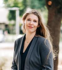
Alyssa Griffin
Assistant Professor, Earth and Planetary Sciences
"Marine sediments in a changing climate: Unlocking the past and preparing for the future"
The various spheres of the Earth system interact through complex physical, biological, and chemical processes that operate over vast spatial and temporal scales. Few aspects of the Earth system are immune to changes resulting from anthropogenic carbon emissions which continue to profoundly shape the nature and habitability of our planet. Marine sediments are the ultimate sink of anthropogenic carbon emissions on geologic timescales and a fundamental control of atmospheric CO2, ocean chemistry, and in turn, Earth’s climate. Many inferences about changes in global climate and seawater chemistry over Earth’s history rely on interpretations of the geochemical signatures contained in marine sediments. Marine sediments are also inextricably linked to present-day marine resources and central to several proposed climate mitigation strategies. Understanding biogeochemical processes related to marine sediments can improve our ability to interpret Earth’s climate through time, respond to present climatic changes, and effectively prepare for the future consequences of climate change.
In this talk, I will discuss my ongoing research at the nexus of marine sediment biogeochemistry and Earth’s changing climate. I will outline my research in the context of unresolved and emerging questions related to carbon cycling in marine sediments across broad spatiotemporal scales. Additionally, I will highlight how studying marine sediments contributes to building coastal resiliency, co-developing mutually beneficial academic partnerships, and elevating the role of the earth science community in addressing the unfolding climate crisis.
Watch a recording of Dr. Griffin's talk on our YouTube channel.
Victoria Keeton
Assistant Professor, Betty Irene Moore School of Nursing

“Social determinants of stress and health in Latinx mothers and children in the U.S.”
November 1, 2023–In this presentation, Dr. Keeton summarizes current evidence around social needs and stressors experienced by many Latinx families in the U.S., and how they may influence the physiology and emotional health of mothers/caregivers and their children. She presents the results of her research in this area, including associations between social needs and hair cortisol concentration as a potential biomarker for chronic stress, and physiologic synchrony in mother-child dyads. Dr. Keeton also discusses the implications of her research findings for clinical practice, health policy, and future research, to address structural drivers of social and health inequities in the Latinx community.
Watch a recording of Dr. Keeton's talk on our YouTube channel.
Luis Diaz-Garcia
Assistant Professor, Viticulture and Enology
Foundations for a modern grape breeding program
May 25, 2023–The UC Davis grape breeding program is shifting its approach to developing new rootstock and scion cultivars. Historically, grape breeding has been largely focused on incorporating single-gene traits related to disease resistance, and fruit and wine quality. However, multi-gene phenotypes such as drought and heat tolerance, fruit yield, soil adaptation, and virus tolerance, are complex and challenging to improve, requiring a gradual increase of small-effect, beneficial alleles. In this talk, I will discuss our efforts to integrate novel phenomics and genomics tools to enhance the breeding of rootstocks and wine grape materials, particularly for complex, quantitative traits that will be crucial for addressing future climate change challenges.
Anna La Torre
Associate Professor, Cell Biology and Human Anatomy
Temporal regulation of neural identity
May 11, 2023–The central nervous system (CNS) contains myriads of different types of neurons. All these different cells are produced from pluripotent neural stem cells during development. Neural stem cells acquire distinct cell identities depending on their spatial position, but they are also influenced by temporal cues to give rise to different cell populations over time. For instance, the progenitors of the cerebral neocortex generate different populations of excitatory projection neurons following a well-known sequence. The Notch signaling pathway plays crucial roles during this process. We have discovered that Notch signaling is essential for neocortical and hippocampal morphogenesis, and for the development of the corpus callosum and choroid plexus. Our data also indicate that in the neocortex, Notch controls projection neuron fate determination through the regulation of two microRNA (miRNA) clusters that include let-7, miR-99a/100, and miR-125b. Our findings collectively suggest that balanced Notch signaling is crucial for telencephalic development and that the interplay between Notch and miRNAs is critical to control neocortical progenitor behaviors and neuron cell fate decisions.
Rose Kagawa
Assistant Professor, Emergency Medicine
Challenges and Promising Practices in Background Check Systems for Firearm Purchases
April 27, 2023–The US prohibits firearm purchase among individuals with specific risk factors, such as convictions for a felony offense. These prohibitions are operationalized using background checks (BC) for firearm purchase. Despite these restrictions, prohibited persons obtain firearms, sometimes with devastating effects. We explore the state of the records and record-keeping practices that underpin BCs for firearm transfers in this mixed-methods, descriptive study. Sources include key informant interviews with individuals working in a leadership capacity with state record-keeping systems, surveys of state systems, and administrative records. We identify potential causes and solutions for challenges relating to missing or incomplete information and delays in resolving ambiguous cases. Examples of potentially causal factors include manual processes (i.e. “human time”) for confirming relationship status in misdemeanor crimes of domestic violence, and discordant practices for processing arrest and court records that result in dispositions that cannot be entered or arrests with no disposition. Potential solutions range from the automation of record sharing across agencies to policies that align prohibiting categories with available records. Background checks are the backbone to many policies designed to prevent firearm harm, yet their effectiveness is subject to the design and operation of the system. We identify numerous actionable ways in which BC practices and policies could be made more robust.
Redefining Health and Wellness: Community-Centered Approaches to Resisting Racism, Sexism, and Transphobia in Medicine
April 20, 2023–Please join the Office of Academic Diversity (OAD) and three of our CAMPOS and CAMPSSAH Faculty Scholars as they discuss their work and the intersections of their research across three public health disciplines. The colloquium will include short presentations from each of the speakers, a panel discussion among speakers moderated by CAMPSSAH Director Zoila Mendoza and an informal discussion.
Chris Hanssmann (Gender, Sexuality, and Women’s Studies), "Epidemiological Biographies: Activist Reconsiderations of Trans and Travesti Health"
This presentation will discuss several community-based investigations conducted in the last two decades in Argentina and the United States that radically reframed conventional epidemiological approaches to understanding and intervening in trans health. These small-scale studies, which I call “epidemiological biographies”, blended narrative and quantitative data to reject the prevailing “health behavior” models that dominated trans health research. Instead, they suggested that criminalization, racialized transphobia, state violence, and immiseration were the primary conditions endangering health and life expectancy in trans and travesti communities. I explain how Buenos Aires- and New York City-based activists mobilized these publications to shift dynamics within public health research and broader political domains. Although activist accounts are frequently portrayed as overly subjective or unscientific, epidemiological biographies took empiricism seriously to highlight what health care providers and researchers were missing in their collective understanding of the needs of trans and travesti communities.
Tiffani Johnson (Emergency Medicine)
Tiffani Johnson’s research portfolio reflects her commitment to improving the quality of care for underserved children. Her interdisciplinary research program is focused on race and racism and its impact on child health. She is currently exploring root causes of inequities in the healthcare and early childhood education settings, including research on racism and bias and its impact on the health and well-being of children.
Adeola Oni-Orisan (Family and Community Medicine), "Community-Centered Approaches to Birth Equity"
High health care spending has not translated into better birth outcomes for Black birthing people in the United States. Structural racism continues to contribute to significant disparities in pregnancy-related mortality and morbidity. How are communities mobilizing and building power to create anti-racist spaces of care for Black birthing people and their families? What barriers do they face? What role, if any, should large research institutions play in this work? This talk will focus on the innovative work of Black birthworkers and Black women-led organization in advancing birth equity.
María Maldonado
Assistant Professor, Plant Biology
Structural Insights Into Respiration Across the Tree of Life
April 13, 2023–Respiration is a key metabolic process driving most life of Earth. Yet, the biochemical and three-dimensional details of the protein complexes that carry out respiration have remained unknown outside of a handful of model organisms, curtailing our understanding of the biodiversity of this process across the tree of life. I will discuss how we can use new biochemical and structural-biology strategies to obtain high-resolution structures of these complexes from low-abundance samples or previously under-studied organisms. The structures we have obtained so far generated new mechanistic hypotheses for respiration and revealed how limited our understanding of this core metabolic process still is.

Wilsaan Joiner
Professor
Neurobiology, Physiology, and Behavior, College of Biological Sciences
Neurology, School of Medicine
Assessing Motor Control Capabilities in Children with Congenital Upper-limb Differences
March 8, 2023–Children with a unilateral congenital below elbow deficiency (UCBED) have one typical upper limb, and one that ends below the elbow, at the proximal level or mid forearm (i.e., lacking a hand). This is an isolated condition, and children with UCBED develop normally. Unlike the majority of adults who usually acquired their limb loss, children with UCBED were born with their limb difference; their affected muscles have never actuated an intact limb. Due to this lack of experience, it has been assumed that their abilities to purposefully modulate muscle activity is limited, subsequently influencing prosthetics designed for this population. To quantify the extent of their control over these residual muscles, we used ultrasound imaging techniques to study 7 children with UCBED (6 congenital, 1 acquired, ages 8-18). We examined the extent children could intentionally and reliably activate the residual muscles to represent distinct hand motions mirrored with their typical hand. We demonstrate that all subjects could enact up to 10 distinct muscle patterns representing different grasp patterns (e.g., power grasp) and could perform these intended movements consistently. We also compared these motor abilities to the unaffected limb and found similar performance across limbs. The results suggest that although participants had never actuated the missing hand they could still distinctively and consistently activate the residual muscles that would perform the intended motor act. This provides insight into how motor control develops in the absence of the normal effector and serves as a guide for designing prostheses that leverage the full extent of these children’s motor control capabilities.
Elva Diaz
Professor, Pharmacology
Chair, Neuroscience Graduate Program
How to change your mind: Molecular mechanisms of brain function underlying learning and memory
February 22, 2023–A key neural mechanism of learning and memory is thought to be activity-dependent changes in AMPA-type glutamatereceptors (AMPAR) levels at synapses, the fundamental units of communication between neurons. Changes in synaptic AMPAR content are driven by long-term potentiation (LTP) and depression (LTD) of synaptic strength. Increases in synaptic strength are mediated by the recruitment of GluA1-containing AMPARs from nearby reserve pools and stabilization at synapses. While significant progress has been made in understanding AMPAR anchoring at synapses, less is known about the mechanisms that govern establishment and maintenance of reserve pools of extrasynaptic AMPARs. Previously, we identified the SynDIG (Synapse Differentiation Induced Gene) family of four genes (SynDIG1-4) encoding brain-specific transmembrane proteins that associate with AMPARs. Here we demonstrate that SynDIG4 (SD4) maintains the reserve pools of GluA1-containing AMPARs that are targeted to synapses during LTP and required for higher order cognitive function such as learning and memory.
Watch Prof. Diaz's presentation on our YouTube channel
Aldrin V. Gomes
Professor and Vice-Chair, Neurobiology, Physiology, and Behavior
Professor, Physiology and Membrane Biology
The Dual Nature of Ibuprofen: Benefits and Precautions
February 8, 2023–Ibuprofen is a commonly used nonsteroidal anti-inflammatory drug (NSAID) that has been widely prescribed for the management of pain and inflammation. Despite its widespread use, both benefits and drawbacks are associated with ibuprofen usage. Long-term use of ibuprofen has been shown to cause adverse effects such as stomach bleeding, increased risk of heart attack or stroke, and kidney damage. Our studies suggest that even moderate amounts of ibuprofen used for shorter periods may have damaging effects on most tissues. A moderate dose of ibuprofen given to mice for 7 days resulted in muscle atrophy, and heart and liver damage. At the molecular level, ibuprofen increased mitochondrial superoxide production which oxidizes several intracellular proteins resulting in numerous signaling pathways being altered. This presentation will provide an overview of the mechanisms, benefits, and risks of ibuprofen use, as well as practical advice for maximizing the benefits while minimizing the risks of ibuprofen usage.
Watch Prof. Gomes's presentation on our YouTube channel
Anya L. Brown
Assistant Professor, Evolution and Ecology, Bodega Marine Laboratory
Ecology of extended phenotypes: stressor effects on coral hosts and their microbes
January 11, 2023–Host-associated microbes are part of a host’s extended phenotype, they can be host-specific, and influence host fitness as a trait of the host. The questions that I ask focus on the implications of variation of traits (including microbes) for organisms, populations and communities. Much of my work is focused on tropical corals and coral reefs. Corals show fantastic amounts of variation: among species, within species and in their physiology, symbionts, and response to stressors. In this seminar I will focus on coral interactions with a biotic stressor, the vermetid gastropod, Ceraesignum maximum, and coral disease and the implications of the intraspecific variation on coral populations and reef communities.
Kenjiro W. Quides
Assistant Professor of Teaching, Microbiology and Molecular Genetics
Experimental Evolution of Rhizobial Symbionts
November 30, 2022–Microbes can dramatically alter the fitness of host organisms, with outcomes ranging from mutualistic to antagonistic depending on the environmental context of the interaction. I use the legume-rhizobium symbiosis to study context dependence of mutualistic interactions. I implement multiple approaches to track shifts in rhizobial population composition including single generation and multi-generation experiments, growth chamber and greenhouse growth systems, and culture-based and molecular methods. I have developed multiple research modules that can be adapted for a Course-based Undergraduate Research Experience (CURE) to foster inclusive excellence in STEM. These modules alone do not make the CURE. Rather, they can be incorporated into a course that is built on student collaboration, sharing of data, and communication of findings.
Esteban Soto
Professor, Medicine & Epidemiology, School of Veterinary Medicine
Emergence of Piscine Lactococcosis in California
November 2, 2022–Aquaculture is the world’s fastest developing food production sector. The industry has experienced rapid growth in recent years as global catches from wild fisheries have reached maximum sustainable yields or declined. Lactococcosis is one of the most serious emerging diseases in fish worldwide. Outbreaks are associated with high mortality rates and significant economic losses, and effective tools for treatment and prevention are limited. A wide range of host species are susceptible, including wild and cultured fish from cold, temperate, and warm fresh or marine water systems. Infections have also been reported in humans and other terrestrial species, making it a disease of concern in aquaculture, conservation, and human and animal health. Historically, cases of piscine lactococcosis have been attributed to the gram-positive bacterium Lactococcus garvieae. Our recent work, however, has revealed that the disease is caused by multiple species - L. garvieae, L. petauri, and L. formosensis that are indistinguishable by conventional diagnostic methods. Lactococcus petauri is arguably the most ruinous emergent pathogen of salmonid fish in the USA. The origin of this pathogen is unknown, but catastrophic outbreaks have occurred in the states of California, Washington, and Arizona causing significant economic losses to conservation programs as well as private aquaculture facilities. A lack of efficacious vaccines, mounting antimicrobial resistance, and limited treatment options result in mortality events >90% in some epizootics. These factors illustrate an urgent and critical need for effective prophylactic measures. This lecture will summarize the research efforts by the UC Davis Aquatic Animal Health laboratory to better understand the epizootiology of piscine lactococcosis and develop safe and effective prophylactic and therapeutic approaches against this disease.
Alvaro Alvarado
Assistant Project Scientist, Kornblum Lab, Semel Institute for Neuroscience and Human Behavior, University of California Los Angeles
Targeting Heterogeneity in Glioblastoma
October 19, 2022–Despite efforts to gain a deeper understanding of its molecular architecture, glioblastoma (GBM) remains uniformly fatal. While recent advances in single cell technology and genomebased subtyping have revealed that GBMs may be parsed into several molecularly distinct categories, this insight has yielded little progress towards extending patient survival. We have developed two separate projects to resolve tumoral heterogeneity: first, we interrogated tumor samples using a pathway-based approach to generate clusters based on overall patterns of enrichment. These data underscore the importance of cell cycle signatures in tumor biology. Second, we created a 28-marker mass cytometry panel composed of signaling markers and lineage markers that have been associated with GBM or cancer stem cells. These studies reveal pathways that are preferentially use by different tumor cells that interact with distinct immune populations. Both studies have the overarching goal of establishing therapeutic efforts to extend patient survival.
Watch the presentation on YouTube
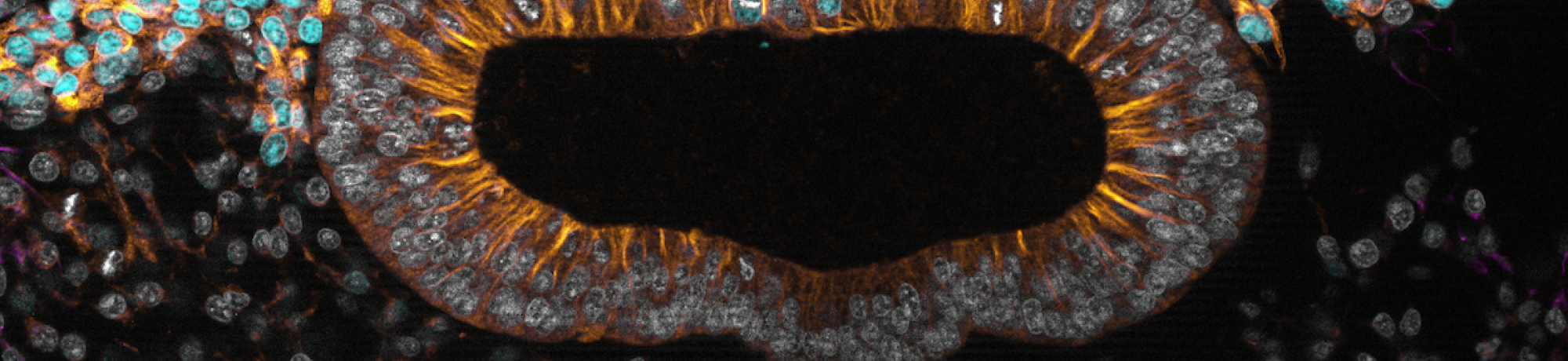
Verónica Martínez-Cerdeño
Professor of Pathology and Laboratory Medicine, School of Medicine
The Pathology of Autism
October 5, 2022–Autism is a neurodevelopmental condition that does not have a cure and for which a distinct pathology has not been found. Current treatments for autism target associated symptoms but not the condition itself. To develop a cure it is imperative that we first determine the brain pathology. We work towards determining pathology, and we have discovered many properties of the brain with autism. Specifically, we found an increase of excitatory pyramidal cells in the upper layers of the cerebral cortex, a decreased number of Chandelier (Ch) cells in the cortex, a decreased number of cartridges, a decreased amount of neurotransmitter receptors in the pyramidal cell axon initial segment -target of Ch cells synapses- and a reduced Ch bouton size -indicating a decrease in the number of synapsis per bouton- in autism. These findings mean that there is an alteration in the excitatory and inhibitory systems in autism.
Watch the recording on our YouTube channel
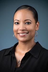
Tiffani Johnson
Assistant Professor, Emergency Medicine, School of Medicine
Using Equity Science to Address Racism as a Public Health Crisis
June 1, 2022–Recent events including police shootings of unarmed Black men, women, and children at the intersection of disparities in the setting of the COVID-19 pandemic have resulted in a long overdue national awakening regarding race and racism in society. In this talk Dr. Johnson will share her ongoing journey using equity science to help address racism as a public health crisis through 4 different phases of research: (1) Identifying racial disparities in pediatric healthcare and outcomes; (2) Investigating and describing racism as a root cause of disparities; (3) Developing and testing interventions aimed at advancing child health equity; (4) Translation- using research as a tool for advocacy.

Alexis Patterson Williams
Associate Professor, School of Education
Sustaining Disciplinary Literacy in Science: A Transformative, Just Model for Teaching the Language of Science
May 26, 2022–The process of developing disciplinary literacy in science is not a neutral act. The acquisition of disciplinary literacy in science has been documented as producing strong, negative emotion in students. In this talk, I will outline the problematic nature of literacy in science, identify specific challenges of science texts as well as highlight strategies for addressing these challenges. Specifically, I identify a culturally sustaining and just approach to teaching science, (W)holistic Science Pedagogy (WSP). It is both a teacher- and student-centered approach of instruction to disrupt patterns and hierarchies. This talk will introduce the WSP commitments within the context of teaching disciplinary literacy in science.
Verónica L. Morales
Assistant Professor, Civil and Environmental Engineering
Groundwater Flow and Mass Transport in Structurally Complex Porous Media
May 18, 2022–This talk will discuss work carried out to understand large-scale behavior as a reflection of the millions of interactions between the moving fluid and the porespaces of the medium. The first part of the talk recasts flow resistance as a graph-theory problem. Through this we learn where and why preferential flow paths form based on information about the pore-structure. The second part of the talk discusses the controls for rock dissolution behavior during CO2 geologic sequestration. This work sheds light on the domino effect that flow, transport and mixing have on reaction outcomes, and presents a predictive model that describes accurately reaction-rate inhibition and reaction-hotspot formation.
Watch the recording on our YouTube channel
Fernanda S. Valdovinos
Assistant Professor, Environmental Science & Policy
Working group to develop theory on plant-herbivore interactions for terrestrial food webs
May 11, 2022–My talk will present the outcomes of our very successful and productive working group at UC Davis in April 11-13 on terrestrial food webs, partially funded by the Jump Start grant. This working group brought together internationally renowned experts on food web ecology, networks, and plant-herbivore interactions. We developed the theoretical framework I have been aiming for the last several years, which we will publish using two outlets: 1) a review article led by me, which will conceptually describe our framework and use some mathematization to illustrate its power, and 2) a research article led by my PhD student, which will implement mathematically one aspect of our novel framework and evaluate how terrestrial vs aquatic ecosystems differ in their food web dynamics. My talk will also describe this novel theoretical framework we developed, which we expect will promote new understanding of how terrestrial food webs respond to global change.
Natalia Caporale
Assistant Professor of Teaching, Neurobiology, Physiology and Behavior
Impacts of Challenging Scientist Stereotypes in Geology Students
May 11, 2022–Geoscience is the least racially diverse of the natural sciences. This is the result of the combination of institutional, social and individual factors leading to the disproportionate attrition of students with identities historically underrepresented in STEM from the field. Students’ sense of belonging, science identity and self-efficacy have been identified as key psychosocial factors for the retention of URM students in STEM. In recent years, interventions that introduce students to non-stereotypical scientists in the context of class material (Scientist Spotlights) have been successfully used in the biological sciences to promote science identity and to disrupt stereotypical perceptions of scientists; changes that may promote retention of URM students. In this study, we developed and then implemented Geoscience-related Scientist Spotlights in the laboratory sections of a Physical Geology course. In this talk I will present the results of this intervention and student feedback on the use of Scientist Spotlights in the classroom and discuss the importance of challenging students' preconceptions of the kinds of people who do science in our efforts to promote diversity in STEM.
Marco S. Messina
UC President's Postdoctoral Research Fellow, College of Chemistry, University of California, Berkeley
Activity-Based Sensing Approaches to Monitor Chemical Analytes in Biological Systems
May 4, 2022–Chemical analytes such as hydrogen peroxide (H2O2), copper, and iron, among others, mediate signaling processes to help maintain homeostasis in complex biological systems. Abnormal changes to intracellular levels of these species are implicated in pathologies such as cancer, neurodegeneration, and autoimmune disorders. The small size and transient nature of these species coupled with the complex environment in which they operate, complicates our ability to monitor them. Our group develops imaging probes that take advantage of activity based sensing (ABS), a process which relies on the inherent differences in chemical reactivity to distinguish between analytes, to generate a fluorescent readout. This presentation will provide an overview of some key tools we have recently developed to (1) monitor transcellular H2O2 signaling and (2) measure relative levels of H2O2, copper, and iron across a human tumor cell line panel using confocal fluorescence microscopy.
Lillian Cruz-Orengo
Assistant Professor, Anatomy, Physiology, and Cell Biology
You Speak with An Accent…Linguistic Profiling in Academia
April 13, 2022–Linguistic profiling is defined by John Baugh as the auditory equivalent of visual racial profiling. It can have devastating consequences for those who are perceived to speak with an undesirable accent or dialect. It is time we talked about linguistic bias and the impact it has on higher education. Also, to consider its significance within a MSI and our efforts towards diversifying the pipeline to academia.
Watch the recording on our YouTube channel
Theanne N. Griffith
Assistant Professor Physiology and Membrane Biology
Illuminating new roles for Nav1.1 in peripheral sensory function
March 9, 2022–Voltage gated sodium channels (Navs) are critical mediators of neuronal excitability and are established therapeutic targets for a variety of nervous system disorders, including epilepsy and pain. Of the nine mammalian isoforms, Nav1.1 is notable for the hundreds of associated disease-causing mutations, which often result in severe brain disease. Nav1.1 is also expressed in the peripheral nervous system; however, due to its prominent role in brain function, the role this channel plays in peripheral neurotransmission remains underexplored. In this presentation, Theanne will discuss recent work from her lab that has identified two new roles for Nav1.1 in peripheral somatosensory processing. She will share evidence for important contributions of Nav1.1 to thermosensation, the ability to detect environmental temperature, and proprioception, the ability to detect limb position for purposeful movement. Collectively, this work has expanded our knowledge of Nav1.1 function in the peripheral nervous system and may provide insight as to how mutation of Nav1.1 affects sensory processing.
Watch the recording on our YouTube channel
Jairo Fúquene Patiño
Assistant Professor, Statistics
Bayesian mixture models, non-local prior formulations and MCMC algorithms
March 2, 2022–In this talk professor Patiño will present the use of Bayesian mixture models in practical settings. He will also discuss Non-local prior alternatives for mixture distributions and Markov Chain Monte Carlo algorithms for posterior inference and model selection. This talk is motivated with real examples.
Jeanette B. Ruiz
Assistant Professor, Communications
Vaccine hesitancy in the age of COVID-19
February 23, 2022–Vaccine hesitancy is not a new phenomenon. However, COVID-19 brought about new challenges for public health officials looking to improve vaccine rates.
Watch the recording on our YouTube channel
Maciel Hernández
Assistant Professor, Human Ecology
A Person-Centered Analysis of Ethnic-Racial Socialization in Adolescence
February 16, 2022–Adolescents commonly contend with and navigate sociocultural pressures and structural inequities such as ethnic-racial discrimination that detract youth from reaching their academic potential. Research studies suggest that ethnic-racial socialization, primarily from parents, and promotion of cultural competence, such as ethnic studies curriculum in schools, provide a positive avenue for youths’ academic competence and well-being. However, youth have simultaneous and proximal interactions with multiple socializing agents, including their peers and school. To expand on current research on ethnic-racial socialization, the purpose of this study was to investigate the existence of ethnic-racial socialization patterns across parent, peer, and school sources and to test whether these patterns relate to academic competence in a sample of ethnically diverse youth.
Special Presentation–Advancing Multicultural Perspectives on Science, Teaching, and Service
February 9, 2022
Theanne Griffith–Science and Stories: A Magnificent Combination
Assistant Professor, Physiology and Membrane Biology
James Anthony Letts–FNL AISES Rocketry: How Engaging with Students Leads to Amazing Opportunities
Assistant Professor, Molecular and Cellular Biology
Jacqueline Barlow
Assistant Professor, Microbiology and Molecular Genetics
Causes and consequences of genome instability in B lymphocytes
January 26, 2022–B cells are highly prone to DNA damage arising from two distinct sources: base modifications made by the enzyme AID (Activation-induced cytidine deaminase) during immunoglobulin gene rearrangement, and replication stress due to their high rate of proliferation in response to antigen stimulus. Thus, primary lymphocytes provide an ideal system to study causes and consequences of planned and unplanned chromosome rearrangements and their role in driving tumorigenesis. In this talk, I will present multiple projects investigating the types of events that arise during normal replication to create damage, as well as the molecular mechanisms driving how “planned” antibody-associated and “unplanned” replication-associated chromosome rearrangements are formed. I will discuss the potential impact of chromosome rearrangements on adaptive immunity and tumorigenesis.
Miriam Nuño
Associate Professor, Biostatistics and Surgery Residence
Math and Public Health: A Focus on Impact
Jan. 19, 2022–Mathematical modeling and statistics are an important part of infectious disease epidemiology and are essential to fight the spread of pathogens. During the COVID-19 pandemic, modeling has played a critical role in assessing the potential impact of interventions, testing efforts, and vaccine. Statistics have been instrumental in facilitating effective communication of COVID-19 risks, assessing uncertainty, and generating projections. COVID-19 revealed major disparities in SAR-CoV-2 infection rates and disease severity in the most vulnerable populations, highlighting the importance of precision public health. In this talk, I will present several projects that highlight unique opportunities of collaboration during this pandemic locally, as well as, state-wide. I will focus on projects that provided actionable insight and guided local public health efforts. I will describe future projects, learned lessons, and opportunities to advance research at the interface of mathematics, statistics, and public health. I will discuss the potential role of mathematics and statistics in addressing health disparities in the COVID-19 pandemic and future outbreaks.
Kristen M George
Assistant Professor, Public Health Sciences
Lifecourse Risk Factors for Cognitive Aging and Impairment
December 1, 2021–Dementia is a complex clinical syndrome that develops over many years and even decades. Researchers are just beginning to recognize that risk factors across the lifecourse contribute to poor cognitive outcomes in late-life. Black and Latinx older adults in the U.S. experience the greatest burden of dementia, and this disparity is likely driven, in part, by greater lifetime exposure to risk factors. This presentation will discuss three studies assessing risk factors at different points in the lifecourse with cognitive outcomes. Kristen will present on the relationship between birth in the Stroke Belt region of the U.S. and late-life cognition among older Black Americans, the association of early adulthood cardiovascular risk factors and late-life cognition in the same cohort of Black Americans, and the relationship between lifetime experiences of discrimination with cognitive decline among a racially/ethnically diverse cohort of individuals ages 90+.
Special Presentation–Advancing Multicultural Perspectives in Research, Teaching, and Science
November 10, 2021
Madeline Nieves-Cintron–Leveraging research, teaching, and service to improve diversity in STEM and serve the community
Assistant Professor, Pharmacology, School of Medicine
Lillian Cruz-Orengo–STEM Building Blocks: Outreach in Veterinary Medicine
Assistant Professor, Anatomy, Physiology & Cell Biology
Verónica Martínez-Cerdeño–The Ventricular Foundation: Science teaching and mentorship
Professor, Pathology and Laboratory Medicine